
Worried about how severe anemia affects your vision? Learn the signs and symptoms of anemic retinopathy, why it causes blurry vision, and how to restore your vision.
Peer-Reviewed By Dr. Ramkrishna Dey, Ophthalmologist, Ocular Health Consultant, Last Review, July 2026.
Your eye doctor found “retinal changes” during a routine check. Your blood report shows low hemoglobin. Now you are searching for answers at midnight, worried about your vision. This fear is common, and it has a name: anemic retinopathy.
As a wellness researcher with over a decade of clinical study experience, I have seen how anemia quietly affects organs beyond the blood. This condition is one such effect, and it is more manageable than you think.
✦ Quick fact: This retinal condition is usually reversible once your anemia gets treated. This guide explains why, and what you should do next.
| Key Takeaways: Anemic Retinopathy * The retinal condition is retinal damage caused by severe anemia reducing oxygen delivery to your eyes. *Common signs include retinal hemorrhages, Roth spots, and cotton wool spots. *Risk rises sharply below hemoglobin 8 to 9 g/dL, especially with low platelets. *Most cases of this condition are reversible once the underlying anemia is treated. *Seek prompt eye care if you have diagnosed anemia and any new vision changes |
Quick Answer: What Is Anemic Retinopathy?
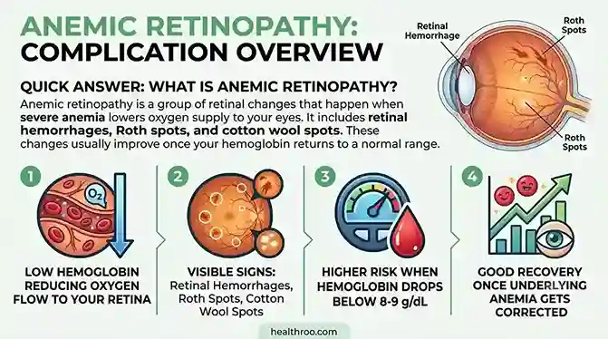
Anemic retinopathy is a group of retinal changes that happen when severe anemia lowers oxygen supply to your eyes. It includes retinal hemorrhages, Roth spots, and cotton wool spots. These changes usually improve once your hemoglobin returns to a normal range.
In short, this eye condition involves:
- Low hemoglobin reducing oxygen flow to your retina
- Visible signs like hemorrhages, Roth spots, and cotton wool spots
- Higher risk when hemoglobin drops below 8 to 9 g/dL
- Good recovery once the underlying anemia gets corrected
What Is Anemic Retinopathy? A Patient-Friendly Explanation
Most medical articles jump straight to clinical terms. Let us slow down first. You deserve a plain answer before the science.
Retina Basics: What You Are Actually Looking At
Your retina is a thin, light-sensitive layer of tissue located at the back of your eye. It captures light and converts it into electrical signals, which are sent to your brain, allowing you to see. This tissue works hard, all day, every day. It needs a steady oxygen supply to function well.
The retina has one of the highest oxygen demands in your entire body. Its blood vessels are thin and delicate. When your blood carries less oxygen, your retina feels the impact early. This is the root cause behind the retinal condition.

Why Anemic Retinopathy Occurs
Several clinical studies say severe anemia lowers your blood’s oxygen-carrying capacity. Your retina, being oxygen-hungry, often shows the first visible signs of this shortage. That is why an eye exam sometimes reveals anemia before a routine blood test does.
How Anemia Affects the Retina: The Science of Anemic Retinopathy
Understanding the mechanism helps you make sense of your diagnosis. Let us break it down simply.
Retinal Hypoxia Due to Low Hemoglobin
Here is the chain reaction behind this condition. Low hemoglobin reduces oxygen-carrying capacity. This starves retinal tissue of oxygen, a state called hypoxia. Hypoxia weakens capillary walls. Weak walls leak blood and fluid. This leakage shows up as hemorrhages on your fundus exam.
The Role of Thrombocytopenia in Anemic Retinopathy
Many patients with severe anemia also have low platelet counts, known as thrombocytopenia. Platelets help seal damaged blood vessels. Without enough platelets, tiny retinal injuries bleed more easily. This combination sharply raises your risk for the condition. Clinical data links anemia with thrombocytopenia to a much higher rate of retinal findings than anemia alone.
Hypoxic vs Ischemic Retinal Damage
Anemic retinopathy differs from diabetic retinopathy in an important way. Diabetic retinopathy happens due to blocked blood vessels, called ischemia. This retinal condition happens due to poor oxygen-carrying capacity, called hypoxia, even when vessels remain open. This distinction matters for diagnosis and treatment. Keep this difference in mind as we compare both conditions later in this guide.

Types of Anemia That Cause Anemic Retinopathy
From the different literature reviewed stated that not every anemia carries equal risk for your eyes. The cause and severity both matter.
By Mechanism
Doctors classify anemia into three broad groups:
- Hypoproliferative anemia, where your bone marrow makes too few red blood cells
- Hemolytic or blood-loss anemia, where red blood cells break down or you lose blood
- Ineffective erythropoiesis, where red blood cell production itself is abnormal
Iron Deficiency Anemia and Anemic Retinopathy
Iron deficiency anemia is the most common trigger for this eye condition, especially across South Asian populations. India’s National Family Health Survey (NFHS-5) reports that anemia affects a large share of women and children nationwide. This makes iron deficiency anemia a leading, preventable cause of this complication in our region.
Megaloblastic Anemia: Vitamin B12 and Folate Deficiency
Vitamin B12 deficiency, also called pernicious anemia, can independently damage retinal capillaries. Folate deficiency works through a similar pathway. Both reduce healthy red blood cell production, lowering oxygen delivery to your retina.
Aplastic Anemia and Sickle Cell Retinopathy
Aplastic anemia, where your bone marrow fails to produce enough blood cells, can also trigger the retinal condition. Sickle cell retinopathy deserves a separate mention. It stems from blocked blood vessels caused by misshapen red blood cells, not from oxygen-carrying capacity alone. Doctors treat it as a distinct condition, though it can overlap with this condition in severe cases.
Anemia From Leukemia and Blood Disorders
Leukemia and related blood disorders can cause a condition called leukemic retinopathy. It shares features with the condition but often includes additional findings like white blood cell infiltration. Your doctor will use blood counts to tell these conditions apart.
Anemic Retinopathy Symptoms You Should Not Ignore
Recognising symptoms early helps you seek timely care.
Common Symptoms of Anemic Retinopathy
Watch for these signs:
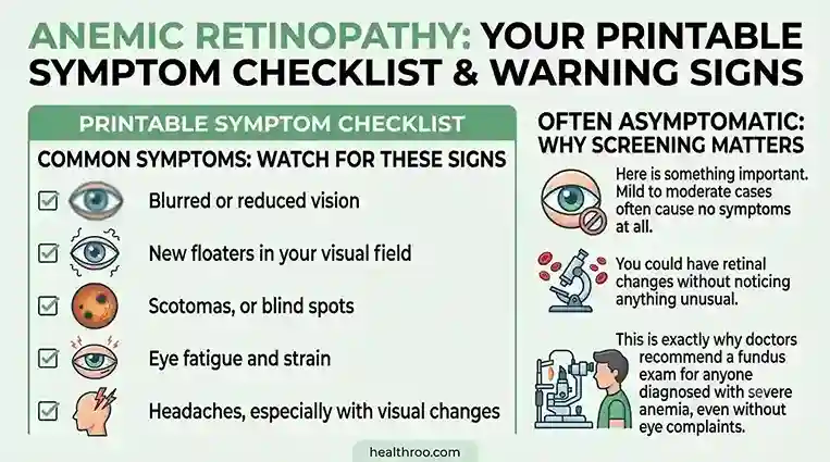
- Blurred or reduced vision
- New floaters in your visual field
- Scotomas, or blind spots
- Eye fatigue and strain
- Headaches, especially with visual changes
Read More Related Issue: Burning sensation in eyes
Often Asymptomatic: Why Screening Matters
Here is something important. Mild to moderate cases often cause no symptoms at all. You could have retinal changes without noticing anything unusual. This is exactly why doctors recommend a fundus exam for anyone diagnosed with severe anemia, even without eye complaints.
✦ Expert insight:
Ocular symptoms linked to these retinal changes can start even before hemoglobin drops, once ferritin falls below 30 ng/mL. Low ferritin, not hemoglobin alone, can signal early risk. This pre-anemic window is often missed in routine checkups.
Signs Your Eye Doctor Looks For in Anemic Retinopathy
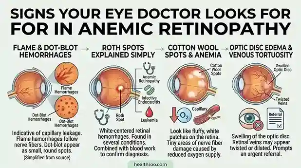
During a fundus exam, your doctor checks for specific patterns linked to this complication.
Flame and Dot-Blot Hemorrhages
Flame hemorrhages look like streaks along nerve fibers. Dot-blot hemorrhages appear as small, round spots. Both types are common findings in the retinal condition and indicate capillary leakage.
Roth Spots Explained Simply
Roth spots are white-centered retinal hemorrhages. They commonly appear in this condition, but they are not exclusive to it. Doctors also see Roth spots in infective endocarditis and leukemia. Your doctor will combine this finding with your blood work to confirm the diagnosis.
Cotton Wool Spots and Anemia
Cotton wool spots look like fluffy, white patches on the retina. They represent tiny areas of nerve fiber damage caused by reduced oxygen supply, a hallmark of the condition.
Read More Related Issue: Photophobia
Optic Disc Edema and Venous Tortuosity
In more severe cases of this retinal condition, the optic disc can swell. Retinal veins may also appear twisted or dilated. These findings often prompt an urgent referral to a retina specialist.
Read More Related Post: Iron Deficiency Eye Floaters
Who Is Most at Risk of Anemic Retinopathy?
Your hemoglobin level plays a central role in predicting this risk.
Hemoglobin Thresholds and Retinal Risk
| Hemoglobin or Parameter | Reported Risk Association |
| Hb below 8 to 9 g/dL | Meaningful rise in retinal changes; around 85.8% sensitivity for this eye condition at Hb below 8.95 g/dL |
| Hb below 6 g/dL | Sharply increased risk of retinal hemorrhage |
| Hematocrit below 30.5% | Around 80% sensitivity for this complication |
| Anemia with thrombocytopenia | Retinopathy reported in up to 28% of anemic patients, rising to 38% with low platelets |
✦ Practical rule:
If your hemoglobin falls below 9 g/dL and you notice vision changes, request a dilated fundus exam without delay. Early detection of retinal conditions can prevent serious complications and help preserve your vision.
Other Risk Factors for Anemic Retinopathy
Beyond hemoglobin levels, several groups face higher risk:
- People with chronic illness, including kidney disease and cancer
- Pregnant women with severe anemia
- Older adults with long-standing nutritional gaps
- Adolescents with heavy, unmanaged menstrual blood loss
Read More Related Issue: Dry eyes and iron deficiency
Anemic Retinopathy in Adolescents: A Growing Concern
This is an area we care deeply about at Healthroo, given our work in adolescent counseling. Teenage girls with heavy or prolonged periods can develop iron deficiency severe enough to affect their eyes, making early recognition and treatment essential. Families and schools rarely connect period-related blood loss to eye symptoms like blurred vision or floaters.
Simple anemia screening in schools, paired with basic eye symptom check-ins, can help detect this condition early, allowing timely treatment and reducing the risk of vision complications. India’s adolescent anemia burden remains high according to NFHS-5 data, which makes school-level screening a practical, low-cost solution worth pursuing.
When Should You See an Eye Doctor? Anemic Retinopathy Decision Guide
Knowing when to act matters as much as knowing the symptoms.
Red-Flag Symptoms
Seek care promptly if you notice:
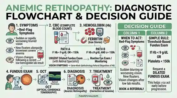
- Sudden or rapidly worsening blurred vision
- New floaters alongside known severe anemia
- Vision changes following a recent low-hemoglobin lab result
Threshold-Based Fundus Exam Guidance
Here is a simple rule to follow. If your hemoglobin is below 9 g/dL, or your platelet count is below 150,000, request a dilated fundus exam. If you notice any vision symptoms at any hemoglobin level, book a same-week ophthalmology referral. This stepwise approach helps you act before the condition progresses.
How Is Anemic Retinopathy Diagnosed?
Diagnosis combines an eye exam with targeted blood work.
Eye Examination and Dilated Fundus Exam
Your doctor will dilate your pupils using eye drops, allowing a clear view of your retina and optic nerve. This allows a clear view of your retina. The exam is quick and painless, though your vision may blur temporarily afterward.
OCT, OCT-A, and Fundus Photography
Optical coherence tomography, or OCT, creates detailed cross-section images of your retina. OCT-A maps blood flow within retinal vessels. Fundus photography documents visible changes over time. These tools help your doctor track this retinal condition as your anemia improves.
Fluorescein Angiography
This test uses a special dye to highlight blood flow in your retina. Doctors reserve it for complex or unclear cases of these retinal changes.
Blood Work: CBC, Ferritin, B12, Platelets
A complete blood count, ferritin level, vitamin B12 level, and platelet count together confirm the cause of this eye condition. Ferritin testing deserves special attention. It can reveal risk even before your hemoglobin drops, since low ferritin often precedes full-blown anemia.
Severity Levels of Anemic Retinopathy
Doctors classify this complication into three severity levels:
- Mild: Isolated findings, usually without symptoms
- Moderate: Multiple hemorrhages or cotton wool spots, with mild visual symptoms possible
- Severe: Extensive hemorrhage, disc swelling, or macular involvement, with noticeable vision change
Medical Disclaimer
This article on anemic retinopathy is for general education only. It does not replace professional medical advice, diagnosis, or treatment. Always consult a qualified ophthalmologist or hematologist for concerns about anemia or vision changes. Healthroo does not endorse self-diagnosis or self-treatment of anemic retinopathy based on this content alone. If you experience sudden vision changes, seek immediate medical attention.
Anemic Retinopathy Treatment: Complete Roadmap
Treatment follows a clear, ordered path.
Step 1: Treat the Underlying Anemia
This is the foundation of the retinal condition treatment:
- Oral or intravenous iron therapy to restore iron levels and correct iron deficiency.
- Vitamin B12 and folate replacement for megaloblastic anemia
- Blood transfusion when anemia is severe or symptomatic
- Hematology-directed treatment when leukemia causes the anemia
Step 2: Ophthalmology Follow-Up
Most retinal findings in this condition are monitored rather than treated directly, as they usually improve once the underlying anemia is corrected. As your hemoglobin improves, the hemorrhages and spots typically resolve on their own.
Step 3: Hematology Referral for Underlying Cause
If your anemia has no obvious cause, or if a marrow disorder is suspected, your doctor will refer you to a hematologist. Correcting the root cause is the real fix for the condition, not just treating the eye findings.
Is Anemic Retinopathy Reversible? Vision Recovery and Prognosis
Yes, and here is how. Most cases of this retinal condition improve fully once the underlying anemia gets corrected.
Recovery Timeline
Retinal changes often begin improving within a few weeks of hemoglobin normalizing. Full recovery timing varies by person, depending on how severe the anemia was and how quickly treatment started.
Predictors of Poor Outcomes
A few factors can slow or limit recovery:
- Very severe anemia at the time of diagnosis
- Concurrent thrombocytopenia
- Macular involvement
- Delayed treatment of the underlying cause
Permanent Damage Risk
Permanent visual impairment from these retinal changes is uncommon. It mainly occurs with macular involvement or significant treatment delay. Most patients recover their vision fully once their anemia gets treated.
Possible Complications of Anemic Retinopathy
While rare, this eye condition can lead to:
- Vitreous hemorrhage
- Macular involvement affecting central vision
- Retinal vascular occlusion, in rare instances
- Permanent visual impairment, though uncommon
Anemic Retinopathy vs Other Retinopathies: Comparison Table
This complication shares features with other retinal conditions, but key differences set it apart.
| Feature | Anemic Retinopathy | Diabetic Retinopathy | Hypertensive Retinopathy |
| Primary mechanism | Hypoxia from low hemoglobin | Vascular ischemia or blockage | Vessel wall damage from high blood pressure |
| Key findings | Roth spots, flame hemorrhages, cotton wool spots | Microaneurysms, new vessel growth | Arteriolar narrowing, AV nicking |
| Reversibility | Often fully reversible | Managed, not typically reversed | Improves with blood pressure control |
| Underlying trigger | Severe anemia | Chronic high blood sugar | Chronic high blood pressure |
Leukemic retinopathy and retinal vein occlusion can also mimic this condition. A blood count review helps your doctor tell these conditions apart with confidence.
Real Patient Cases: Anemic Retinopathy in Practice
The following cases are illustrative, built from common clinical presentation patterns of the condition. They are not records of specific named patients.
Case One: The Teenager With Heavy Periods
A 17-year-old girl came in with three months of fatigue and occasional blurred vision. Her periods had grown heavier over the past year. Her hemoglobin measured 6.8 g/dL, with ferritin critically low. A dilated fundus exam revealed several flame hemorrhages and two Roth spots in both eyes. This pattern is typical of anemic retinopathy caused by severe iron deficiency.
Her care team started iron therapy, along with gynecology support for her heavy periods. Within six weeks, her hemoglobin rose above 10 g/dL. Her follow-up fundus exam showed the hemorrhages resolving. Her vision returned to normal within two months. This case reflects how quickly this condition can improve once its root cause gets addressed, especially in young patients.
Case Two: The Older Adult With Chronic Kidney Disease
A 61-year-old man with long-standing kidney disease reported new floaters and mild vision blur. His hemoglobin was 7.2 g/dL, and his platelet count was mildly reduced. His fundus exam showed cotton wool spots and mild optic disc swelling, both consistent with anemic retinopathy linked to his chronic condition.
His nephrologist adjusted his anemia management, including erythropoiesis-stimulating therapy and iron supplementation. His hemoglobin improved gradually over three months. His retinal findings resolved in step with his blood counts, though his ophthalmologist continued monitoring given his kidney disease. This case shows how this eye condition in chronic illness often needs coordinated care between multiple specialists.
Prevention Tips for Anemic Retinopathy
Preventing this complication starts with preventing severe anemia:
- Eat a balanced, iron and B12-rich diet, including dals, leafy greens, and dairy, with supplementation when your doctor recommends it
- Get anemia diagnosed early through routine blood work
- Schedule regular eye exams if you belong to a high-risk group, including pregnant women, adolescents with heavy periods, and people with chronic illness
Frequently Asked Questions About Anemic Retinopathy
Can iron deficiency cause anemic retinopathy?
Yes, iron deficiency anemia is the most common trigger for anemic retinopathy. When hemoglobin falls low enough, your retina does not receive adequate oxygen, leading to hemorrhages, Roth spots, and cotton wool spots. Correcting iron deficiency early through diet or supplements reduces your chances of developing anemic retinopathy. If you already have low iron levels, ask your doctor about a fundus exam, even without visual symptoms.
Is anemic retinopathy an emergency?
Anemic retinopathy is not usually a medical emergency, but it should never be ignored. If you experience sudden vision changes along with known severe anemia, seek prompt ophthalmology review. Doctors need to confirm whether your findings represent anemic retinopathy or a more urgent retinal condition. Quick evaluation ensures you receive the right treatment early and prevents unnecessary anxiety about your eyesight and overall health.
Is retinal hemorrhage from anemic retinopathy reversible?
Yes, in most cases, retinal hemorrhages linked to anemic retinopathy resolve fully once your underlying anemia gets corrected. Recovery often starts within weeks of your hemoglobin improving. Severity at diagnosis, platelet count, and how quickly you start treatment all influence your healing timeline. Regular follow-up fundus exams help your doctor confirm that your anemic retinopathy is improving steadily as expected, week by week.
Can low hemoglobin really affect eyesight?
Yes, hemoglobin below roughly 8 to 9 g/dL is strongly associated with anemic retinopathy and measurable visual symptoms like blurred vision and floaters. Your retina needs a steady oxygen supply, and low hemoglobin directly reduces that supply. This is why doctors recommend eye exams for patients with severe anemia, even when vision seems normal, since anemic retinopathy can develop silently in its early stages.
Can children or teenagers develop anemic retinopathy?
Yes, adolescents with significant iron deficiency, particularly from heavy menstrual bleeding, can develop anemic retinopathy. This age group is often overlooked in eye health discussions, yet the risk is real. Parents and school health programs should watch for fatigue, pale skin, and vision complaints together. Early identification and treatment of anemia in teenagers can prevent anemic retinopathy from progressing to more severe stages.
How long does recovery from anemic retinopathy take?
Retinal changes in anemic retinopathy often begin improving within a few weeks of hemoglobin normalizing, though full recovery time varies between patients. Factors like initial severity, presence of thrombocytopenia, and treatment speed all affect your timeline. Most people see significant improvement within two to three months. Your doctor will schedule follow-up fundus exams to confirm that your anemic retinopathy is resolving on schedule
Can anemia cause eye floaters?
Yes, floaters can be an early symptom of anemic retinopathy, even when other symptoms are absent. Floaters happen when small hemorrhages or debris appear within your retina or vitreous, disrupting your visual field slightly. If you notice new floaters and also have a history of anemia, mention both to your doctor. This combination often prompts a fundus exam to check for anemic retinopathy.
Does vitamin B12 deficiency cause anemic retinopathy?
Yes, megaloblastic anemia from vitamin B12 or folate deficiency can independently lead to anemic retinopathy through the same hypoxic mechanism seen in iron deficiency anemia. Vitamin B12 deficiency, also called pernicious anemia, reduces healthy red blood cell production, lowering oxygen delivery to your retina. Correcting B12 levels through injections or supplements often resolves the retinal findings linked to anemic retinopathy within a few months.
Reference:
PMC (NIH) – Determinants for Anemic Retinopathy
PubMed – Anemic Retinopathy (classic reference)
PubMed – Prevalence of Retinopathy in Anemia/Thrombocytopenia (Carraro et al.)
PubMed – Anemic Retinopathy Secondary to Malnutrition, Case Report
NCBI StatPearls – Roth Spots
NCBI StatPearls – Retinal Hemorrhage
Cleveland Clinic – Iron-Deficiency Anemia
Mayo Clinic – Iron Deficiency Anemia: Symptoms & Causes
WHO – Anaemia Fact Sheet
IIPS/MoHFW – NFHS-5 India Report
Ministry of Health & Family Welfare – NFHS-5 Fact Sheet
Author Bio
Sajal Samanta holds an MSc in Anthropology and works as an adolescent counselor, bringing a human-centered lens to health writing. With over 15 years of experience in RCT-based clinical research, Sajal founded Healthroo.com to translate complex medical evidence into clear, actionable guidance for Indian readers. His work focuses on adolescent wellness, nutritional deficiency, and integrative health, grounded in both clinical data and cultural context.









5 thoughts on “Anemic Retinopathy: Can Severe Anemia Damage Your Eyes?”